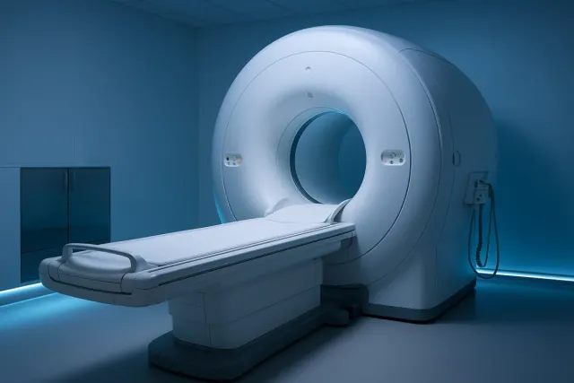
Magnetic Resonance Imaging (MRI) of the Breast
3 dakika okuma süresi
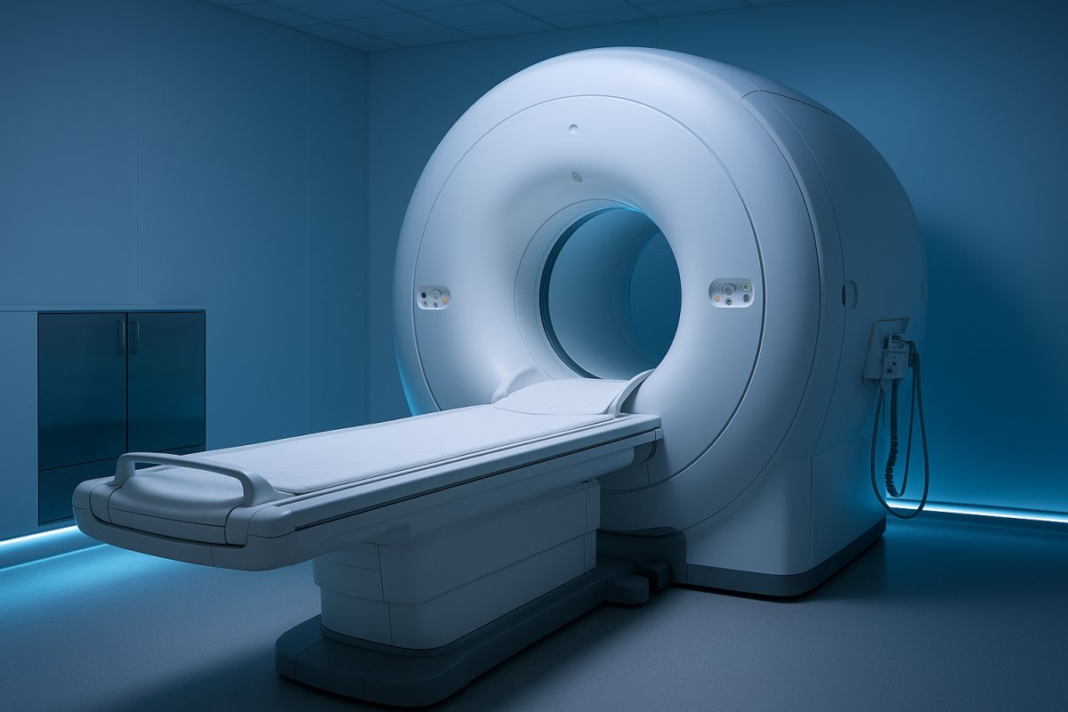
Magnetic resonance imaging (MRI) of the breast produces detailed images using a strong magnetic field and radio waves, without ionising radiation. It is the most sensitive breast imaging method, but it is not a routine screening tool for every woman: it is used in defined situations, complementing mammography and ultrasound.
When Is Breast MRI Requested?
- High-risk screening: annual imaging in addition to mammography for BRCA carriers and women at comparably high risk
- Newly diagnosed breast cancer: assessing disease extent, additional foci and the opposite breast
- Monitoring treatment response: measuring response in patients receiving chemotherapy before surgery
- Clarifying a suspicious finding: when mammography and ultrasound are inconclusive
- Implant assessment: evaluating the integrity of silicone implants
- Unknown primary: when cancer is found in an axillary node but no breast focus is identified
How the Examination Is Performed
- The patient lies face down with the breasts positioned inside a dedicated coil.
- In most examinations a contrast agent (gadolinium) is given intravenously to assess the blood supply characteristics of tissue.
- The scan takes roughly 20-40 minutes and it is important to stay still throughout.
- The scanner is loud; headphones or ear plugs are provided.
Why Timing Matters
In premenopausal women breast tissue changes hormonally through the menstrual cycle, which affects contrast enhancement and can lead to misinterpretation. For this reason the scan is usually preferred in the second week after the start of menstruation.
Strengths and Limitations
Strengths: no ionising radiation, high sensitivity even in dense breast tissue, and accuracy in showing the extent of disease.
Limitations: benign changes can also enhance, so false positive findings are more frequent, which may lead to additional tests and biopsies. It is also less effective than mammography for microcalcifications, costs more, and can be difficult for patients with claustrophobia.
MRI-Guided Biopsy
For suspicious findings seen only on MRI, with no correlate on mammography or ultrasound, sampling can be performed under MRI guidance so that tissue is taken safely from the lesion.
Frequently Asked Questions
Should every woman have a breast MRI?
No. It is not a routine screening tool and is requested for high-risk groups and specific medical indications. Unnecessary use can lead to false positives and avoidable biopsies.
Is the contrast agent harmful?
Gadolinium-based agents are generally well tolerated. Impaired kidney function, pregnancy and previous allergic reactions must be reported.
I am claustrophobic, what can I do?
Tell the team in advance. Wide-bore scanners, breathing and relaxation techniques, or sedation under medical supervision may make the scan possible.
Does MRI replace mammography?
No. They provide different information and MRI does not replace mammography for microcalcifications. The most accurate result comes from assessing the methods together.
I have implants, can I have an MRI?
Yes; MRI is the most sensitive method for assessing implant integrity, and this examination can often be performed without contrast.
Consultation
To find out whether a breast MRI is needed or to review an existing report, you are welcome to get in touch.