Fibroadenoma Treatment with Vacuum Excision Method (Seno Rx Encor)
2 min read
Fibroadenomas are very common benign breast masses in women. They are most common between the ages of 20 and 40. When first detected, they are followed up at six-month intervals for up to two years. The reason for this follow-up is to prevent malignant tumors that resemble fibroadenomas from being mistaken for fibroadenomas. If a fibroadenoma is rapidly growing, painful, or causing the patient discomfort due to a palpable mass, it can be treated. A biopsy is essential to confirm the diagnosis before fibroadenoma treatment.
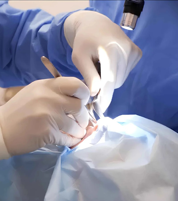
With advancing technology, vacuum biopsy needles are now used instead of standard needles used in pre-operative biopsies, allowing complete removal of the fibroadenoma. This new vacuum-assisted biopsy technique allows all breast masses up to 4 cm, especially fibroadenomas, to be treated in a painless, bloodless procedure lasting just a few minutes.
This method, which is more affordable than surgical treatments, has not yet become widely available in our country because it is currently performed by very few interventional radiologists. We successfully apply this new treatment method to patients who present to our clinic for non-surgical fibroadenoma treatment.
The vacuum excision method allows for the removal of fibroadenomas up to 3-4 cm in pieces. Vacuum excision can remove masses up to 4 cm in diameter in as little as 10-20 minutes. The needle puncture heals without leaving any scars. You can return to your daily life immediately after treatment.
Furthermore, vacuum biopsy can also be used to remove microcalcifications. Ultrasound or mammography guidance is required for vacuum excision of microcalcifications. First, microcalcifications are visualized on ultrasound, and if visible, they can be easily removed with vacuum excision. Microcalcifications not visible on ultrasound can be removed under stereotactic mammography guidance.